13.) Gram staining differentiates Gram-negative bacteria from Gram-positive bacteria, how they are carried out in the laboratory, and discuss the mechanism of action of the stain.
13.) Gram staining differentiates Gram-negative bacteria from Gram-positive bacteria, how they are carried out in the laboratory, and discuss the mechanism of action of the stain.
A.)
Bacteria are a large group of minute, unicellular, microscopic organisms, which have been classified as prokaryotic cells, as they lack a true nucleus. These microscopic organisms comprise a simple physical structure, including cell wall, capsule, DNA, pili, flagellum, cytoplasm and ribosomes.
Bacteria can be gram-positive or gram-negative depending upon the staining methods. Let us have a detailed look at the difference between the two types of bacteria.
Gram Staining
This technique was proposed by Christian Gram to distinguish the two types of bacteria based on the difference in their cell wall structures. The gram-positive bacteria retain the crystal violet dye, which is because of their thick layer of peptidoglycan in the cell wall.
This process distinguishes bacteria by identifying peptidoglycan that is found in the cell wall of the gram-positive bacteria. A very small layer of peptidoglycan is dissolved in gram-negative bacteria when alcohol is added.
Gram-Positive and Gram-Negative Bacteria – Overview
The gram-positive bacteria retain the crystal violet colour and stain purple whereas the gram-negative bacteria lose crystal violet and stain red. Thus, the two types of bacteria are distinguished by gram staining.
Gram-negative bacteria are more resistant to antibodies because their cell wall is impenetrable.
Gram-positive and gram-negative bacteria are classified based on their ability to hold the gram stain. The gram-negative bacteria are stained by a counterstain such as safranin, and they are de-stained because of the alcohol wash. Hence under a microscope, they are noticeably pink in colour. Gram-positive bacteria, on the other hand, retains the gram stain and show a visible violet colour upon the application of mordant (iodine) and ethanol (alcohol).
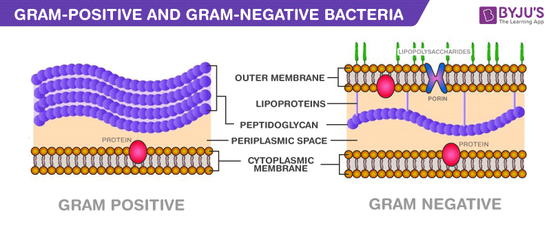
Gram-positive bacteria constitute a cell wall, which is mainly composed of multiple layers of peptidoglycan that forms a rigid and thick structure. Its cell wall additionally has teichoic acids and phosphate. The teichoic acids present in the gram-positive bacteria are of two types – the lipoteichoic acid and the teichoic wall acid.
In gram-negative bacteria, the cell wall is made up of an outer membrane and several layers of peptidoglycan. The outer membrane is composed of lipoproteins, phospholipids, and LPS. The peptidoglycan stays intact to lipoproteins of the outer membrane that is located in the fluid-like periplasm between the plasma membrane and the outer membrane. The periplasm is contained with proteins and degrading enzymes which assist in transporting molecules.
The cell walls of the gram-negative bacteria, unlike the gram-positive, lacks the teichoic acid. Due to the presence of porins, the outer membrane is permeable to nutrition, water, food, iron, etc.
Difference between Gram-Positive and Gram-Negative Bacteria – Key Points
- The cell wall of gram-positive bacteria is composed of thick layers peptidoglycan.
- The cell wall of gram-negative bacteria is composed of thin layers of peptidoglycan.
- In the gram staining procedure, gram-positive cells retain the purple coloured stain.
- In the gram staining procedure, gram-negative cells do not retain the purple coloured stain.
- Gram-positive bacteria produce exotoxins.
- Gram-negative bacteria produce endotoxins.
Difference between Gram-Positive and Gram-Negative Bacteria
Following are the important differences between gram-positive and gram-negative bacteria:

Difference between Gram-Positive and Gram-Negative Bacteria
| Gram-Positive bacteria | Gram-Negative bacteria |
| Cell Wall | |
| A single-layered, smooth cell wall | A double-layered, wavy cell-wall |
| Cell Wall thickness | |
| The thickness of the cell wall is 20 to 80 nanometres | The thickness of the cell wall is 8 to 10 nanometres |
| Peptidoglycan Layer | |
| It is a thick layer/ also can be multilayered | It is a thin layer/ often single-layered. |
| Teichoic acids | |
| Presence of teichoic acids | Absence of teichoic acids |
| Outer membrane | |
| The outer membrane is absent | The outer membrane is present (mostly) |
| Porins | |
| Absent | Occurs in Outer Membrane |
| Mesosome | |
| It is more prominent. | It is less prominent. |
| Morphology | |
| Cocci or spore-forming rods | Non-spore forming rods. |
| Flagella Structure | |
| Two rings in basal body | Four rings in basal body |
| Lipid content | |
| Very low | 20 to 30% |
| Lipopolysaccharide | |
| Absent | Present |
| Toxin Produced | |
| Exotoxins | Endotoxins or Exotoxins |
| Resistance to Antibiotic | |
| More susceptible | More resistant |
| Examples | |
| Staphylococcus, Streptococcus, etc. | Escherichia, Salmonella, etc. |
| Gram Staining | |
| These bacteria retain the crystal violet colour even after they are washed with acetone or alcohol and appear as purple-coloured when examined under the microscope after gram staining. | These bacteria do not retain the stain colour even after they are washed with acetone or alcohol and appear as pink-coloured when examined under the microscope after gram staining. |
Gram staining procedure in laboratory.
At the lab, a medical laboratory scientist smears or spreads the sample on glass microscope slides. These slides are known as smears. They then apply a series of stains to the smear to perform a Gram stain.
The Gram staining process includes four basic steps, including:
- Applying a primary stain (crystal violet).
- Adding a mordant (Gram’s iodine).
- Rapid decolorization with ethanol, acetone or a mixture of both.
- Counterstaining with safranin.
Examining the Gram stain
The medical laboratory scientist then categorizes any bacteria that may be present by color and shape during the microscopic evaluation:
- Color: Typically, bacteria that are gram-positive appear purple to blue, and bacteria that are Gram-negative appear pink to red.
- Shape: The most common shapes include round (cocci) or rod-shaped (bacilli).
The medical laboratory scientist also looks for additional characteristics of the sample by observing the groupings of the bacteria on the slide. Examples include:
- Cocci that are present singly, in pairs, in groups of four, in clusters or in chains.
- Bacilli that are thick, thin, short, long or have enlarged spores on one end.
- If bacteria are present within white blood cells.
The medical laboratory scientist then puts together a report and sends it to your healthcare provider.
What do the results of a Gram stain mean?
Gram stain test results reveal one of two categories: a negative Gram stain or a positive Gram stain. This is not to be confused with gram-negative bacteria or gram-positive bacteria.
Negative Gram stain
If your test result reveals a negative Gram stain or “no organism seen,” it usually means that there are too few bacteria present to be able to be seen using the Gram stain method. Bacteria might still be detected by culture if a culture is performed on the specimen.
Positive Gram stain
If your test result reveals a positive Gram stain, it means that bacteria were present in your sample. If your result is positive, it usually includes information about what kind of organism was present on the sample slide, including:
- Type of bacteria: Gram-positive or gram-negative.
- Shape of bacteria: Round (cocci) or rods (bacilli).
- Other bacteria characteristics: Size, relative quantity (number) and/or arrangement of the bacteria, if applicable.
- Other cells: Whether there are bacteria present within other cells (intracellular) and if there’s a presence of red blood cells or white blood cells.
- Fungi: Gram stains can check for the presence of fungi in the form of yeasts or molds. You may need further testing to identify the specific type.
This information, along with signs and symptoms and other clinical findings, will help your healthcare provider determine which treatment may be most effective, sometimes before bacteria culture results are available.
Gram staining is a differential staining technique.
Bacteria are classified as Gram positive and Gram negative based on the differences in their cell wall composition. Gram negative bacterial cell wall consists of an outer membrane which is absent in Gram positive bacteria.
- In Gram’s staining the primary stain used is crystal violet which stains all the cells purple irrespective of the composition of cell wall.
- Next, mordant fixes the crystal violet to the cell wall.
- Further, 95% alcohol (decolourizer) increases the porosity of cell walls which contain LipoPolySaccharide layer (LPS). When the poro
Comments
Post a Comment