Difference between Prokaryotic cell and Eukaryotic cell, Differences Between Plant Cell and Animal Cell ,Fluorescence microscope principle ,ray diagram and instrumentation
krish creations
1.) Difference between Prokaryotic cell and Eukaryotic cell.
Prokaryotic cells are primitive cells mainly found in the unicellular organisms, and they do not nucleus, instead they contain a fragment of DNA, and the organelles are not bounded by the membranes, where as the eukaryotic cells are found in all types of multicellular organisms such as plant and animal cells and they contains nucleus and membrane bound organelles.
| Prokaryotic cells | Eukaryotic cells |
| This cells are always unicellular | Eukaryotic cells are present as either unicellular or multicellular. |
| The size of cell is generally range from 0.2 micrometers to 2.0 micrometers in diameter | Eukaryotic cells range from 10 to 100 micrometers in diameter. |
| In prokaryotic cells, the cell wall is present and it is very complex in nature. | Eukaryotic cells have cell walls very rarely, if present they have simple chemical nature. |
| In this cells true nucleus absent, instead nucleotide is present | True nucleus is present. |
| DNA is arranged in circular shape | DNA is linear in shape |
| In prokaryotic cells, cytoplasm is present, but it is lacking in most cell organelles. | In eukaryotic cells, it consists of both cytoplasm and organelles, both are present. |
| Mitochondria is absent | Mitochondria is present and it is a powerhouse of cells. |
| Ribosomes are present, and they are small in size and shape is spherical | Ribosomes are present but they are comparatively large and linear in shape. |
| Endoplasmic reticulum and lysosomes and centromere’s all are absent | Endoplasmic reticulum and lysosomes and centromere’s all are present. |
| Plasmids are commonly found in prokaryotes. | Plasmids are very rarely found in eukaryotes |
| Cell division occur through binary fission | Cell division occur through mitosis |
| Flagella is small in size | If flagella are large in size. |
| In this cells only asexual reproduction occurs. | Both sexual and asexual reproduction occurs. |
| Bacteria and Archaea are examples. | Plant and animal cells are examples. |
Note: So prokaryotes are primitive cells compared to eukaryotes and in prokaryotes both transcription and translation occurred as couple, which means that translation begins during the time of synthesis of mRNA, while in eukaryotic cells, mRNA produce in nucleus and enters the cytoplasm where translation begins freshly.
2.) Differences Between Plant Cell and Animal Cell

Diagram showing the Difference between Plant cell and Animal cell
Diagram showing the Difference between Plant cell and Animal cell
As stated above, both plant and animal cells share a few common cell organelles, as both are eukaryotes. The function of all these organelles is said to be very much similar. However, there are major differences between plant and animal cells.
The major differences between the plant cell and animal cell are mentioned below:
| Plant Cell | Animal Cell |
| Cell Shape | |
| Square or rectangular in shape | Irregular or round in shape |
| Cell Wall | |
| Present | Absent |
| Plasma/Cell Membrane | |
| Present | Present |
| Endoplasmic Reticulum | |
| Present | Present |
| Nucleus | |
| Present and lies on one side of the cell | Present and lies in the centre of the cell |
| Lysosomes | |
| Present but are very rare | Present |
| Golgi Apparatus | |
| Present | Present |
| Cytoplasm | |
| Present | Present |
| Ribosomes | |
| Present | Present |
| Plastids | |
| Present | Absent |
| Vacuoles | |
| Few large or a single, centrally positioned vacuole | Usually small and numerous |
| Cilia | |
| Absent | Present in most of the animal cells |
| Mitochondria | |
| Present but fewer in number | Present and are numerous |
| Mode of Nutrition | |
| Primarily autotrophic | Heterotrophic |
Conclusion
Both plant and animal cells comprise membrane-bound organelles, such as endoplasmic reticulum, mitochondria, the nucleus, Golgi apparatus, and lysosomes. The plant cell can also be larger than the animal cell. The normal range of the animal cell varies from about 10 – 30 micrometres and that of plant cell range between 10 – 100 micrometres.
3.) Fluorescence microscope principle ,ray diagram and instrumentation

The main components of a fluorescence microscope are:
Light source: A high-intensity light source such as a mercury or xenon arc lamp.
Excitation filter: A filter that selectively transmits light at the wavelength that excites the fluorescent molecules in the sample.
Sample: The sample contains fluorescent molecules that emit light at a longer wavelength when excited.
Objective lens: Collects the emitted light and magnifies it to form an image.
Emission filter: A filter that selectively transmits the fluorescence emitted by the sample and blocks the excitation light.
Detector: Collects the emitted fluorescence and converts it into an electronic signal that generates the image.
Fluorescence microscopy can also include additional components such as a dichroic mirror to separate the excitation and emission wavelengths, and confocal optics to increase resolution.
Application :
- Fluorescence microscopes are used in biological research to observe specific molecules and structures within cells and tissues.
- Fluorescence microscopes are used in medical diagnosis to detect fluorescent markers in tissues or cells.
- Fluorescence microscopes are used in neuroscience research to study brain function and development.
- Fluorescence microscopes are used in materials science to study the properties of materials at the nanoscale.
- Fluorescence microscopes are used in environmental science to study environmental samples.
- Fluorescence microscopes are used in forensic science to detect and analyze trace evidence.
Advantages:
High sensitivity: Fluorescence microscopes are highly sensitive and can detect very small amounts of fluorescent signals.
Specificity: Fluorescent dyes can be used to specifically label certain structures, cells or molecules of interest, making it possible to visualize and study them in detail.
Live-cell imaging: Fluorescence microscopy can be used to observe living cells in real time, allowing researchers to study dynamic processes and cell behavior.
Multicolor imaging: Fluorescence microscopy can be used to image multiple fluorescent probes with different colors simultaneously, which allows for the study of multiple targets at once.
High spatial resolution: Fluorescence microscopy can achieve high spatial resolution, allowing researchers to visualize and study structures at the subcellular level.
Disadvantages:
Cost: Fluorescence microscopes are expensive, and their cost can be a barrier to entry for many researchers.
Photobleaching: Fluorescent dyes can degrade over time due to exposure to light, leading to loss of signal over time.
Phototoxicity: Exposure to fluorescent light can damage cells and cause phototoxicity, which can affect cell function and viability.
Background fluorescence: Fluorescent dyes can sometimes generate unwanted background signals, which can interfere with the detection of the target signal.
Limited penetration: Fluorescent light can only penetrate a certain depth into tissues, which can limit the use of fluorescence microscopy for deep tissue imaging.
4.) bright field microscope principle ,ray diagram and
instrumentation.
Bright field microscopes consist of the following basic components:
Light source: typically a halogen bulb that provides bright, uniform illumination to the specimen.
Condenser lens: collects and focuses the light from the light source onto the specimen.
Specimen stage: holds the specimen in place for observation.
Objective lens: magnifies the image of the specimen.
Ocular lens: further magnifies the image projected by the objective lens for observation by the human eye or a camera.
Diaphragm: regulates the amount of light passing through the specimen and into the objective lens.
Focus knobs: used to adjust the focus of the specimen for a clear and sharp image.
Modern bright field microscopes may also have additional features such as digital imaging capabilities, automated stage movement, and fluorescence microscopy attachments.
- Bright field microscopes are widely used in biological research to study the structure and function of cells, tissues, and organisms.
- Bright field microscopes are used in medical settings to diagnose diseases and conditions by examining bodily fluids.
- Bright field microscopes are used in industrial settings to inspect the quality of materials and products.
- Bright field microscopes are used in environmental science to study organisms in their natural habitats.
- Bright field microscopes are commonly used in education to teach students about the microscopic world and study specimens.
Advantages:
Simplicity: Bright field microscopes are relatively simple and easy to use, making them ideal for beginners or students.
Low cost: Bright field microscopes are generally less expensive than more advanced microscopes such as fluorescence microscopes.
High resolution: Bright field microscopes can achieve high resolution, allowing for detailed observation of small structures.
Wide field of view: Bright field microscopes have a wide field of view, making it possible to observe large specimens or multiple specimens at once.
Compatibility with stains: Bright field microscopy can be used in conjunction with various stains, allowing for the visualization of specific structures or cells.
Disadvantages:
Lack of contrast: Bright field microscopy can lack contrast, making it difficult to distinguish between different structures or cells.
Limited visualization of transparent specimens: Bright field microscopy is not well-suited for transparent specimens, as the light passes through them without generating significant contrast.
Limited depth of field: Bright field microscopes have a limited depth of field, which can make it difficult to observe structures that are not in the same focal plane.
Limited sensitivity: Bright field microscopy is not very sensitive to low levels of light or fluorescence, which can make it difficult to detect faint signals.
Inability to observe living cells: Bright field microscopy cannot be used to observe living cells in real-time, as the bright light can damage or kill the cells.
![Ray diagrams for (a) Bright-field imaging using direct beam and (b) dark-field imaging using a specific off-axis scattered beam.(reproduced from [Petr06])](https://www.researchgate.net/profile/Haritha-Nukala/publication/47715467/figure/fig7/AS:307416148267019@1450305129783/Ray-diagrams-for-a-Bright-field-imaging-using-direct-beam-and-b-dark-field-imaging.png) |
Ray diagrams for (a) Bright-field imaging using direct beam and (b) dark-field imaging using a specific off-axis scattered beam |
5.) Dark field microscope principle ,ray diagram and
instrumentation.
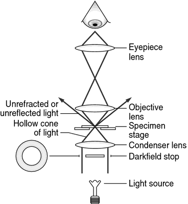
The instrumentation of a dark field microscope includes:
Light source: A light source, such as an LED or halogen lamp, that produces a cone of light that is directed at the specimen.
Condenser: A special condenser that collects and focuses the cone of light at an oblique angle, causing it to pass through the specimen and create a dark background.
Objective lens: A high-powered objective lens that captures and magnifies the scattered light from the specimen.
Eyepiece: An eyepiece that further magnifies the image and allows the viewer to see the specimen.
Iris diaphragm: An iris diaphragm that controls the amount of light entering the condenser.
Specimen holder: A stage or holder that holds the specimen in place and allows for precise positioning.
Adjustment knobs: Knobs that adjust the focus and position of the objective lens to obtain a clear image.
- Dark field microscopy is used to observe live microorganisms, aiding in disease diagnosis.
- Dark field microscopy is used to examine blood cells for disease detection.
- Dark field microscopy is used to observe and test the structure and behavior of materials.
- Dark field microscopy is used to visualize and characterize nanoparticles and other nanoscale structures for various applications.
- Dark field microscopy is used in marine biology to observe and study microscopic organisms, providing insights into marine ecosystems.


Comments
Post a Comment